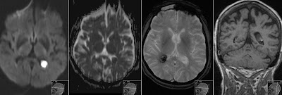
Subdural Hemorrhage in Neonate
T1 weighted images from 3T MRI showing Subdural Hemorrhage (SDH) in the left occipital region in this one week old neonate. Note high signal of SDH on T1 sequence without contrast due to Methemoglobin. There is also Caput Succadeneum.
You can also see SDH on transversal T2 as low signal (black), as high signal (white) on transversal Inversion Recovery (IR) sequence, followed by sagittal T2 and coronal T1. Especially on sagittal T2 note Subarachnoid Space compressed by SDH. Such perinatal SDH are not so rare, however are difficult to find on Ultrasound as they are located in periphery and often do not cause large displacement.


