Dilated Perivascular Spaces in the Brain
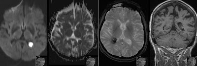
Above images (tra T2, tra FLAIR, cor T2 and sag T1) show enlarged Perivascular Spaces (PVS) in the right frontal lobe of the elderly patient with dementia. Note that signal follows that of the CSF (see FLAIR). Moreover note moderate general cortical atrophy.
PVS, also known as Virchow-Robin spaces are considered normal finding although perhaps in the future we should know more about the exact patophysiology and possible relations till dementia and other conditions.


