Posts

Showing posts with the label Fun

X-mas presents from AI

Image
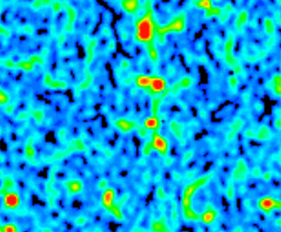
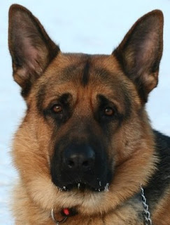
I asked ChatGPT-4: "Can you make me an image of a simulated Lego set of an MRI Scanner. Show it in foreground with its box behind."  This is what I got:  This image was generated by AI called Dall-E that is subdivision of ChatGPT. It looks pretty impressive! It would certainly be a great present for X-mas! I was inspired to make this prompt after seeing the image below that i found on X, that also looks like generated by AI.  I think capabilities of AI in the field of Radiology are pretty impressive!  But honestly I was even more impressed when I asked ChatGPT to spot the difference between two images I took in my garden. It did the great job and this is quite promising example of how can AI be used in Radiology. See the images for yourself first and spot the difference:  And now look at result of AI analysis: As a fun task I asked AI to make me an image of a cat sitting on top of aquarium - something I'm used to see in my office. Here is what I got: I think it'...

AI vs Human

Image
  As a funny post here example of counting spinal levels by AI (Artificial Intelligence - purple) vs Human (me - orange). :)

Music Staff on MRI Lumbar Spine - fun

Image
As a new hobby project I recently took on some digital piano and synthesizer training. Part of it is to learn to read the notes. And somehow I keep on seeing five lines of staff even om MRI images :) ! This localizer incidentally even shows the grand staff . :)  You can see a little annular tear in a place of previous disk herniation.  The numbering of lumbar levels is often a matter of discussion. Previous exam has called the lowest lumbar vertebra as L5. I used the same numbering but in fact the vertebra that is called L5 on this image is a transitional vertebra that is partially sacral vertebra - so it could be called S1. I made a note on my report for the clinician that in case of operation it is important to correlate the level of operation with fluoroscopy .  For correct numbering of spinal lumbar levels I look for proximal parts of ribs 12 on sagittal images and count from what I assume as L5 upwards. When I have the whole spine or CT, especially with coronal im...

Snus on CT

Image
Unfortunately it is very popular in Sweden to use small tobacco bags placed under the upper lip - locally known as Snus . When I saw for the first time a nice Swedish lady placing such tobacco bag under her lip during a meeting - I was shocked with disgust. Note snus bag under the lip as "incidental finding" on the above CT. This case expands "my collection" of various objects found in patient's mouth. You might check my previous:  Chewing Gum on CT and  Candy Under the Tongue on CT . 

Chewing Gum on CT

Image
On the light side of radiology imaging one can notice a chewing gum located above the tongue on this unenhanced CT. Rather easy "diagnosis" however worth recognition as in some cases such foreign bodies can be confusing. See also my previous post about the candy:  Candy Under the Tongue on CT .

Just Looking!

Image
Amusing T2 slice from 3T MRI scanner showing right sided fixed position of the lenses. Perhaps our patient saw something interesting in the scanner? :) For more fun radiology images check older posts in Fun label.

Blue Eyes

Image
Surface reconstruction image from CTA showing both ophthalmic arteries and intra-orbital structures (shaded in blue). Nice for viewing anatomy and from aesthetic perspective. There is also some diagnostic value in such reconstructions as it seems easier to find vascular anomalies and present those to clinical doctors. Oh, and images look much better in motion due to 3D effect.

Not Similar

Image
The above screen-grab is a result of my Google Image search for MRI in Parkinson with selection to "show similar images" (that is to the first image above). Rather surprising and unexpected result! If computed assisted radiology is going to function like Google Image Search engine - than we are long way before computers are replacing humans at the radiology workstations.

Peace and Love from C2

Image
Today a bit of positive humor in normal anatomy. Note a peace sign in the odontoid process at the upper image that corresponds with closed ossification centers. Lower image shows a heart sign that is a normal configuration of the spinous process of the second cervical vertebra (C2) - same vertebra as above. Example of "positive reading" of the radiology images. ;-)

iPhone on CT Scanogram

Image
I always recommend checking the Scanogram on every CT. It can reveal prior operations, foreign bodies - that can be of importance for MRI screening. One can also appreciate patient's general constitution. Sometimes as in this case it can reveal what type of mobile device the patient is using and that he needs glasses to read from it ;).

Candy Under the Tongue on CT

Image
Above CTA shows hyperdense (450 HU) oval structure under the tongue. Imaging is with iv contrast so one could wonder if this is a hyperdense or enhancing lesion? However it is really just a candy that the patient has had under her tongue during examination. Also note the artifacts from dental fillings. Sometimes is worth to angulate the head to avoid those artifact in the regions of interest. There are even some articles about foreign bodies in oral cavity: McDermott M - What's in your mouth? The CT appearance of comestible intraoral foreign bodies. Towbin AJ - The CT appearance of intraoral chewing gum.

Neonatal MRI Faces

Image
This is not a Thai Buddist temple guard. Above image is a coronal T2 of the neonate. Note normal teeth buds in the maxilla. Also low signal from lenses. This is not a cute monkey. Not even Homer Simpson. Above image is a coronal T1 of the neonate. Note normal fat in chicks.

Color art in CT Perfusion images of the brain.

Image
Here some nice colors of the CT Perfusion images of the brain. Showing: Mean Transit Time (MTT) - how fast the blood flows through the brain, Cerebral Blood Flow - how much blood flows in a specific period (ml of blood/100g tissue/min), and Cerebral Blood Volume - how much blood is in a volume of tissue. Excellent pdf article on CT Perfusion of the Brain from Martin Wiesmann is here:  CT Perfusion of the Brain - Martin Wiesmann  
Image
Hi, Welcome to my blog.