Posts

Showing posts with the label Head Neck

Anomalous ossicle between laminae C2 and C3

Image
  There are many congenital anomalies of the spine. One rare is this one spotted as incidental finding on MRI of the Cervical Spine of a 64 years old patient. Reporting radiologist pointed skeletal anomaly in form of an ossicle between laminae of C2 and C3 on the right side causing slight impression on the posterior part of the dural sack, recommending further analysis with CT of the Cervical Spine. The CT has confirmed what looks like an anomalous ossicle between laminae C2 and C3. The ossicle has its own cortex and there are some depressions in the laminae due to its presence.  Similar anomalous ossicle is described in T. Keats - Atlas of Normal Roentgen Variants 7th edition, Figure 3-69, page 215. 
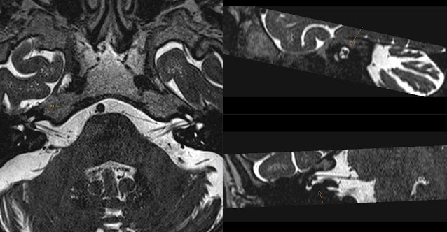
Small Vestibular Schwannoma

Image
Remember my old post about anatomy of the Internal Acoustic Canal (ICA). I mentioned that to me it looks like " Monkey Looking at Cerebellum " in cross section. Mouth of the monkey being Vestibular Nerve and upper eye being 7th nerve (Facial Nerve) (7-UP!) and lower eye being Cochlear Nerve.  Today I came across this exam showing a Small Vestibular Schwannoma (VS) deep in the Internal Acoustic Canal.  Note that the VS is located in the "mouth" of the monkey.  This is a section just next to the VS showing a bit "sad face of the monkey". :(

Orbital Cavernous Hemangioma MRI

Image
Note well defined strongly enhancing mass located medially in the right orbit. This extraconal mass has high signal on T2. It has not changed over time. This is Orbital Cavernous Hemangioma . If we would perform dynamic contrast scan it would show graduate enhancement. Here is the mass nicely depicted on T2, T1 FS with contrast and on CT. Case is complementary to my previous case .

Paraganglioma - Glomus Jugulare

Image
Note intensively enhancing tumour in the foramen jugulare that expands the foramen and with characteristic "salt & pepper" appearance. It extends along the jugular vein and has irregular border. Finding represents Glomus Jugulare Paraganglioma .

Carotidynia

Image
50 years old patient presented with right sided neck pain in the carotid region. Initial CT Angiography and MR Angiography have not shown signs of dissection. However a soft tissue mass around carotid bifurcation on the right side was noted on the initial CT. MR has confirmed poorly defined contrast enhancing mass around carotid bifurcation on the right. Note high signal on the first (fat suppressed) T2 sequences, contrast enhancing on fat suppressed T1 (third image) and better delineation on conventional T2 (last image). Combining clinical and radiological findings the concluded diagnosis is of Carotidynia . This is an idiopathic neck pain syndrome that is mostly diagnosed clinically and reacts well to therapy. Radiological MRI studies show inflammatory tissue in the affected region. Interesting articles on Carotidynia: Bradford S - MR Imaging of Patients with Carotidynia K.E.A. van der Bogt - Carotidynia: A Rare Diagnosis in Vascular Surgery Practice

Orbital Cavernous Hemangioma

Image
Contrast enhanced CT showing intraorbital mass in the orbita apex with a  well defined margin . Mass displaces inferior rectus muscle (arrowheads). It is not a process that extends from the optic nerve sheath neither from the orbit muscle. It does not have infiltrative appearance. There is contrast enhancement but on native CT (not shown) it also presents some high and low attenuation. Mass represents  Orbital Cavernous Hemangioma .

Ossification of the Posterior Longitudinal Ligament

Image
46 years old presents with neurological symptoms from upper extremities. MRI shows significant cervical spinal stenosis that is due to  Ossification of the Posterior Longitudinal Ligament (OPLL) . This is know to progress even after laminoplasty. Surgeons are interested in staging of OPLL. Therefore we perform CT that helps in evaluation of calcifications. Staging of OPLL depends on extension of calcifications, if those are interrupted, continuous or mixed. In this case calcifications are interrupted - type B. This has prognostic value concerning the progress of the disease after laminoplasty. See also staging and overview in this excellent article: Yoshiharu Kawaguchi - Progression of Ossification of the Posterior Longitudinal Ligament Following en Bloc Cervical Laminoplasty

Incisive Canal Cyst on CT

Image
Incidental, benign finding on CT showing cyst in the trajectory of the incisive canal representing Incisive Canal Cyst , aka Nasopalatine Duct Cyst . Clues here are well defined borders and location. Developmental anomaly.

Fishbone in Larynx

Image
Unenhanced CT showing large (about 30mm) fishbone dorsally in the larynx . It is located supraglottic. Its oblique trajectory can be seen in the coronary plane. Hyperdense opacity of the fishbone allows for good depiction with CT. There is also surrounding edema around this foreign body. Initial laryngoscopy failed to reveal the fishbone as it was deeply sunk in soft tissues.

Pneumoparotid

Image
Incidental finding on a trauma CT scan showing air in ducts of the right parotid gland. Note also air in the main  parotid duct . This is known as Pneumoparotid . It is caused by periodic increased air pressure in the oral cavity. It can be seen in people playing wind instruments, after dental procedures, coughing and other activities involving forceful exhalation.

Titanium Mesh

Image
Surface renderings of the CT showing Titanium Mesh used in cranioplasty to cover extensive defect in the right temporal bone. Defect was due to craniectomy after intracranial hemorrhage following trauma. Also note old zygomatic bone fractures.

External Auditory Canal Fracture

Image
Fall on mandible with blood from both external auditory canals. CT shows bilateral comminute fracture of the bony parts of external auditory canals (arrows). Fractures are not apparent at first glance, however clue here is emphysema - air in soft tissues medially to mandibular condyles (arrow heads). Mechanism of this trauma is impact of mandibular condyles against anterior parts of the bony external auditory canals. Lesson here is to look carefully at any abnormal air in soft tissues of trauma patients and ask yourself a question: Where does this air come from?

MRI - Metal Screening

Image
Before every MRI we screen our patients for possible metal foreign bodies. Those can be implanted due to prior medical procedures as well as accidental. One of the specific inquiry concerns possible metal foreign bodies in the orbit. Sometimes patients work with metal and indicate possibility of being exposed to small projectile metal particles. In such case we check prior CT examinations for possible metal foreign bodies. Note above case of a small piece of metal located medially and superiorly in the left orbit. Here metal seems not to be attached to the globe. However due to risk of metal motion in strong magnetic field of the MRI scanner in a case like this we ask for ophthalmologist consultation. One should review scanogram as well as thin slices of the CT - considering size of foreign body and slice thickness. In case we do not have prior CT we order x-ray of the orbits.

Optic Nerve Sheath Meningioma

Image
Contrast enhanced CT showing well defined, homogeneous intra orbital tumor. There is relation of the tumor with optic sheath and rather deep location in the orbital apex. This is most likely an  Optic Nerve Sheath Meningioma .

Struma

Image
3D rendering of the CT Angiography showing nicely a large struma displacing trachea without deforming trachea lumen. Eye Candy.

Monkey Looking at Cerebellum

Image
When looking at the contents of the Internal Acoustic Canal it always reminds me of a face of the " Monkey Looking at Cerebellum ". So I developed a simple way to remember its contents. Note the position of the oblique sagittal T2 slice that is indicated on the topogram and its angle that is shown on the right. You recognize cerebellum easily on the left image. Long arrow on the first image shows Vestibular Nerve entering Internal Acoustic Meatus. Oblique sagittal T2 sequence through the Internal Acoustic Canal showing it's contents: 02:00 - Superior Vestibular Nerve 04:00 - Inferior Vestibular Nerve 08:00 - Cochlear Nerve 10:00 - Facial Nerve. In other words the "mouth" of our "monkey" has a V letter shape since it is a "smiling monkey" and V represents Vestibular Nerve (Superior and Inferior). The upper "eye" is the Facial Nerve (7Up!) and the lower "eye" is the Cochlear Nerve. We can follow the Cochlear Nerve to t...

Le Fort Type 1 Fracture

Image
Note horizontal fracture Le Fort Type 1 involving maxillary sinus walls bilateraly. This type of the fracture causes instability of the alveolar process of the maxilla. Note fractured maxillary sinus walls, pterygoid process, blood levels in both maxillary sinus and extensive soft tissue emphysema. Le Fort fractures are divided in levels: Type 1 - below nose, Type 2 - above nose, Type 3 - through the orbits. Above schema is from Wikipedia that nicely presents types of Le Fort fractures.

Occipital Dural Arteriovenous Fistula

Image
Patient resents with left sided pulsatile tinnitus. CTA reveals pathologically increased arterial circulation of the Occipital Artery and Posterior Auricular Artery branches (arrows). Also noted is arterial contrast intensity in the left Sigmoid Sinus and Internal Jugular Vein (arrowheads). Finding is consistent with Dural Arteriovenous Fistula (DAVF) from the Occipital Artery circulation. Note increased size and number of the branches from the Occipital Artery circulation.

Carotis Segments

Image
Images from MR Angiography (MRA) with Time of Flight (ToF) source sequence on the left and reconstructed Maximum Intensity Projection (MIP) showing stenoses in the Internal Carotid Artery (ICA). This patient has a stent in the distal carotid artery that beginns in petrous and ends in cavernous segment. Presence of the stent can influence images of this ToF a non-contrast MRA. You can see stenosis (arrow). In such case I would recommend CTA. I would like to use this case for a short reminder of the terminology concerning ICA segments. It is easier in CTA since we have reference anatomy points but just in case of the above MIP the typical curves can help. Here are the names of the Internal Carotid Artery Segments: C1 = cervical C2 = petrous C3 = lacerum C4 = cavernous C5 = clinoidal C6 = ophthalmic C7 = communicating Location of the markers corresponds with beginning of the segments (except for C1). As we see our patient has stenosis in the C3 and C4 segments. Also note that th...

Persistent Hypoglossal Artery

Image
Large Persistent Hypoglossal Artery (PHA) is seen traversing the hypoglossal canal. Note origin of this large vessel (Persistent Hypoglossal Artery marked with arrow) that is from distal Internal Carotid Artery (ICA) (arrow head) and located posteriorly to it. 3D reconstructions with mandible partially removed show PHA (arrow) as a large vessel posterior to ICA. Also note how it feeds the basilar artery after exiting from the hypoglossal canal. This case is very interesting not only due to persistent vessel but also that PHA is the only feeding vessel for the basilar artery since both vertebral arteries are hypoplastic and there are no posterior communicating arteries. Interesting article form RadioGraphics: Normal Variants of the Cerebral Circulation at Multidetector CT Angiography - Simon J. Dimmick