Posts

Showing posts with the label Anatomy

Herniated Lumbar Disk MRI

Image
  T2 sequences showing right sided paramedian disk herniation at L5-S1 level compressing right nerve root S1 in the lateral recess. No spinal stenosis. See my YouTube Short Video

Spinal Canal Schwannoma

Image
  A case of a young patient with incidental finding of an intra-dural extra-spinal Schwannoma. Tumor enhances mostly in the periphery, is well defined and shows high signal on T2 sequences. Tumor compresses conus. Note fine outcome on the control study 6 months after operation (on the right) with no residue tumor and fine remodelling of the conus.  See also my YouTube Short . 

Anomalous ossicle between laminae C2 and C3

Image
  There are many congenital anomalies of the spine. One rare is this one spotted as incidental finding on MRI of the Cervical Spine of a 64 years old patient. Reporting radiologist pointed skeletal anomaly in form of an ossicle between laminae of C2 and C3 on the right side causing slight impression on the posterior part of the dural sack, recommending further analysis with CT of the Cervical Spine. The CT has confirmed what looks like an anomalous ossicle between laminae C2 and C3. The ossicle has its own cortex and there are some depressions in the laminae due to its presence.  Similar anomalous ossicle is described in T. Keats - Atlas of Normal Roentgen Variants 7th edition, Figure 3-69, page 215. 

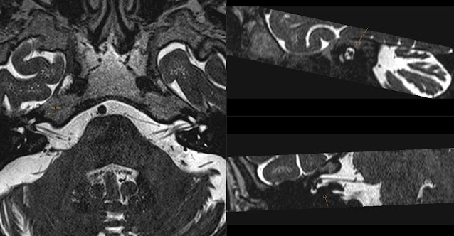
Small Vestibular Schwannoma

Image
Remember my old post about anatomy of the Internal Acoustic Canal (ICA). I mentioned that to me it looks like " Monkey Looking at Cerebellum " in cross section. Mouth of the monkey being Vestibular Nerve and upper eye being 7th nerve (Facial Nerve) (7-UP!) and lower eye being Cochlear Nerve.  Today I came across this exam showing a Small Vestibular Schwannoma (VS) deep in the Internal Acoustic Canal.  Note that the VS is located in the "mouth" of the monkey.  This is a section just next to the VS showing a bit "sad face of the monkey". :(

MRI Tractography - Brain Tumor

Image
Recently when reporting a brain MRI of a patient after tumor operation I came across beautiful functional and MRI Tractography images showing corticospinal tracts relation to the brain tumor in the frontal lobe. The interpretation of this study was that most ventral parts of the corticospinal tracts are located near tumor's dorsal part and the arcuate fasciculus was unaffected. Of course there were more images showing the arcuate fasciculus in more detail. The exam also included functional (fMRI) study showing motoric and speech locations. I'm not expert in this field but I wanted to show you that we are moving closer to using such functional studies in daily practice. 

CT Pelvimetry - Bäckenmätning

Image
Below is a short summary of the protocol for Pelvimetry (Bäckenmätning in Swedish). First a thick MIP is reconstructed on the scanner that covers whole pelvis. A line is drawn on the scanner that is later used for calibration on PACS workstation. Calibration of the previously measured line. On the above MIP the Transversal Inlet Diameter (TransverstingÃ¥ngsmÃ¥tt) is measured as the largest transversal distance in the pelvic inlet. As well as Intertubar Diameter (IntertubaravstÃ¥nd) as distance between  ischial tubers . On sagittal reconstruction measure Sagittal Inlet Diameter (Sagittellt ingÃ¥ngsmÃ¥tt) as shortest distance between posterior part of symphysis pubis and promontorium (S1). As well as Sagittal Outlet Diameter (Sagittellt utgÃ¥ngsmÃ¥tt) as shortest distance between posterior part of symphysis pubis and fist mobile joint between sacrum and coccyx . On axial reconstruction (see topogram for level) measure Interspinal Diameter (Interspinal avstÃ¥n...

Smiling Perivascular Space

Image
Note this enlarged perivascular space in a typical location in the lower parts of the basal ganglia. The "smiling face" represents small vessels.  Contrast enhanced T1 shows the small vessels even better. You can also check my other examples of Perivascular Spaces and Dilated Perivascular Spaces .

Neurocentral Synchondrosis

Image
MRI of the 11 years old child showing symmetric low signal intensity lines on T2 sequences in all vertebrae (arrows). Those are normal growth plates called Neurocentral Synchondrosis  that ossify later in life and are no longer visible in the adult spine. See drawing from Gray's Anatomy figure 102.

Spinal Neurinoma

Image
Patient presented with acute left C2 nerve root neurological symptoms. MR revealed at the level C1-2 a 'dumbbell' shaped intraforaminal mass that was well defined, with high signal on T2, rather homogeneously enhancing, located predominantly extradural and compromising spinal cord significantly. There was also enlargement of the intervertebral foramen without signs of aggressive bone destruction. Radiologic appearance of Spinal Neurinoma . Prompt operation confirmed diagnosis of Schwannoma that was mostly located extradural however with also some intradural component that has been easily evacuated during operation. This case gives opportunity to remind the anatomical location of the spinal nerves. At the level of the cervical spine nerve roots above vertebra have the same number as the vertebra. While in the rest of the spine nerve roots below vertebra have the same number as the vertebra. Therefore nerve root C1 exits above C1 vertebra, C7 root a...

Broca's Infarct

Image
Patient presented with expressive dysphasia . MRI shows acute infarct in Broca's Area in the left inferior frontal gyrus that is responsible for speech production. Note high signal on DWI, low on corresponding ADC as well as signal changes on T2 and coronal FLAIR.

Aneurysm of Persistent Trigeminal Artery

Image
CTA showing large aneurysm at the dorsal upper part of the right Internal Carotid Artery (arrow). There is also an anatomic variant in form of a Persistent Trigeminal Artery (PTA) on the left side which is also showing an aneurysm in its anterior medial part (arrowhead). Note hypoplastic Basilar Artery (long arrow), as another anatomic variant. There are reports in the literature of aneurysms accompanying PTA. This was an incidental finding. Note same findings on the 3D volume reconstructions. (Images are mirrored L-R). Same 3D reconstructions from anterior angle. In this case aneurysms are obvious and easy to find. However it is good to be sure to look for aneurysms in case of this anatomic variant. You might also check my previous post about Persistent Trigeminal Artery . Also, if you like anatomic variants, there is a case of Persistent Hypoglossal Artery .

MCA Territory

Image
Unenhanced CT showing tissue loss after Middle Cerebral Artery MCA infarct involving whole territory of MCA. Note sparring of occipital lobe as well as medial temporal lobe - that belong to Posterior Cerebral Artery PCA territory. Also note medial parts of the frontal lobe that belong to Anterior Cerebral Artery ACA. Above unenhanced CT from the investigation three months prior at acute stage of infarct showing " Dense Vessel Sign " indicating occlusion of the main M1 segment of the MCA on the left side. CT Perfusion study at the time of acute infarct showing: increased Mean Transit Time MTT, decreased Blood Volume BV and decreased Blood Flow BF. Especially the decreased BF - represented by dark blue in the last image indicates infarct that can not be saved. Remember: " Blue is Bad " in CT Perfusion.

AV Hemodialysis Shunt Thrombosis

Image
CTA (Computed Tomography Angiography) showing nice 3D rendering of the AV Hemodialysis Shunt Thrombosis (purple) in the cephalic vein with dilatation of the distal part of the cephalic vein. The Arterio-Venous shunt is open. Source images of the same investigation show that there is partially blood flow around thrombus and dilated distal part of the cephalic vein. Important note: CTA is not the investigation of choice for the suspected hemodialysis shunt thrombosis - Color Doppler Ultrasound is the investigation that should be performed first. This case was investigated in order to provide surgeon with anatomic topography of the anastomosis and in this way replacing angiography.

Anterior Commissure

Image
Why I like Anterior Commissure ? It is a horizontal stripe of tissue connecting hemispheres that is located below frontal horns and anterior and superior to the third ventricle. It is very thin in antero-posterior dimension and very distinct on coronal images. Above two coronal 1.4mm thick T1 contrast enhanced images look identical but are from different examinations of the same patient. Anterior Commissure is helpful in synchronizing those two sequences for comparison e.g. in tumor growth. I also love anatomy drawings. The above is Figure 744 from Henry Gray (1825–1861). Anatomy of the Human Body. 1918. Courtesy Bartleby.com  

Monkey Looking at Cerebellum

Image
When looking at the contents of the Internal Acoustic Canal it always reminds me of a face of the " Monkey Looking at Cerebellum ". So I developed a simple way to remember its contents. Note the position of the oblique sagittal T2 slice that is indicated on the topogram and its angle that is shown on the right. You recognize cerebellum easily on the left image. Long arrow on the first image shows Vestibular Nerve entering Internal Acoustic Meatus. Oblique sagittal T2 sequence through the Internal Acoustic Canal showing it's contents: 02:00 - Superior Vestibular Nerve 04:00 - Inferior Vestibular Nerve 08:00 - Cochlear Nerve 10:00 - Facial Nerve. In other words the "mouth" of our "monkey" has a V letter shape since it is a "smiling monkey" and V represents Vestibular Nerve (Superior and Inferior). The upper "eye" is the Facial Nerve (7Up!) and the lower "eye" is the Cochlear Nerve. We can follow the Cochlear Nerve to t...

Oculomotor Infarct

Image
This 40 years old patient has experienced acute oculomotor nerve paresis. A 1.5T MRI has revealed a micro infarct (about 2mm) in location of the oculomotor nerve nucleus in the mesencephalon. Note high signal on DWI (B1000), low signal on ADC and high on T2 (B0) of the Diffusion Weighted Images (DWI) and coronal FLAIR. This case illustrates how sensitive can DWI be even in small infarcts. Above drawing courtesy of Bartleby . Fig. 710 Anatomy of the Human Body by Henry Gray This is also a great opportunity to remind the location of the oculomotor nerve nucleus in mesencephalon (8'). Note that mesencephalon here is "facing down" compared to MRI. Well seen structures on the MRI are Red Nucleus, Substantia Nigra and Cerebral Aqueduct. Cranial Nerves Nuclei Drawing titled: Brain stem human sagittal section - by Patrick J. Lynch , medical illustrator Courtesy Wikipedia Also note location of the other Cranial Nerves Nuclei in the brainstem that are nicely depicted in th...

Carotis Segments

Image
Images from MR Angiography (MRA) with Time of Flight (ToF) source sequence on the left and reconstructed Maximum Intensity Projection (MIP) showing stenoses in the Internal Carotid Artery (ICA). This patient has a stent in the distal carotid artery that beginns in petrous and ends in cavernous segment. Presence of the stent can influence images of this ToF a non-contrast MRA. You can see stenosis (arrow). In such case I would recommend CTA. I would like to use this case for a short reminder of the terminology concerning ICA segments. It is easier in CTA since we have reference anatomy points but just in case of the above MIP the typical curves can help. Here are the names of the Internal Carotid Artery Segments: C1 = cervical C2 = petrous C3 = lacerum C4 = cavernous C5 = clinoidal C6 = ophthalmic C7 = communicating Location of the markers corresponds with beginning of the segments (except for C1). As we see our patient has stenosis in the C3 and C4 segments. Also note that th...

Persistent Hypoglossal Artery

Image
Large Persistent Hypoglossal Artery (PHA) is seen traversing the hypoglossal canal. Note origin of this large vessel (Persistent Hypoglossal Artery marked with arrow) that is from distal Internal Carotid Artery (ICA) (arrow head) and located posteriorly to it. 3D reconstructions with mandible partially removed show PHA (arrow) as a large vessel posterior to ICA. Also note how it feeds the basilar artery after exiting from the hypoglossal canal. This case is very interesting not only due to persistent vessel but also that PHA is the only feeding vessel for the basilar artery since both vertebral arteries are hypoplastic and there are no posterior communicating arteries. Interesting article form RadioGraphics: Normal Variants of the Cerebral Circulation at Multidetector CT Angiography - Simon J. Dimmick