Posts

Showing posts with the label Spine

The Lasting Imprint of Poliovirus on the Spine and Musculature

Image
  Radiology Case Review: The Lasting Imprint of Poliovirus on the Spine and Musculature While widespread vaccination efforts have made acute poliomyelitis a rare encounter in modern clinical practice, radiologists still frequently observe the chronic, lasting footprints of the virus in adult patients. Today, we are reviewing a fascinating and historically significant MRI case that beautifully illustrates the long-term sequelae of polio on both the central nervous system and the musculoskeletal system. The poliovirus has a notorious affinity for the lower motor neurons, specifically targeting the anterior horn cells of the spinal cord. Decades after the initial infection, the resultant damage presents with a very characteristic set of imaging findings. Let’s break down the key features visible in this patient’s scans. 1. Symmetrical Anterior Signal Changes in the Spinal Cord Looking at the axial T2-weighted images of the cervical spine, the pathophysiology of the virus is strikingly...

Spinal Extradural Arachnoid Cyst (Nabors Type IA)

Image
  Case: Spinal Extradural Arachnoid Cyst (Nabors Type IA) Imaging Modality:   MRI Thoracolumbar Spine   Sequences Provided:   Sagittal T2, Axial T2, Sagittal T1 Radiologic Findings:   The MRI demonstrates a well-circumscribed, unilocular cystic lesion located within the posterior extradural space of the lower thoracic spine, at approximately the T11-T12 level. The lesion follows cerebrospinal fluid (CSF) signal characteristics across all pulse sequences, appearing uniformly hyperintense on T2-weighted images and hypointense on T1-weighted images. Spanning approximately 24 mm in craniocaudal dimension, the cyst exerts focal mass effect upon the thecal sac. It causes significant anterior displacement and compression of the distal spinal cord. The axial T2-weighted sequence confirms its posterolateral extradural position, illustrating the resultant flattening of the dural sac. There are no internal septations, complex features, or solid components visible. Impressi...

Herniated Lumbar Disk MRI

Image
  T2 sequences showing right sided paramedian disk herniation at L5-S1 level compressing right nerve root S1 in the lateral recess. No spinal stenosis. See my YouTube Short Video

Spinal Canal Schwannoma

Image
  A case of a young patient with incidental finding of an intra-dural extra-spinal Schwannoma. Tumor enhances mostly in the periphery, is well defined and shows high signal on T2 sequences. Tumor compresses conus. Note fine outcome on the control study 6 months after operation (on the right) with no residue tumor and fine remodelling of the conus.  See also my YouTube Short . 

Vitamin B12 deficiency related subacute combined degeneration of the spinal cord

Image
  Young patient diagnosed with vitamin B12 deficiency showing classical inverted V sign in the posterior part of the cervical spinal cord that indicates Subacute Combined Degeneration of the spinal cord.  See also my YouTube Short :

Anomalous ossicle between laminae C2 and C3

Image
  There are many congenital anomalies of the spine. One rare is this one spotted as incidental finding on MRI of the Cervical Spine of a 64 years old patient. Reporting radiologist pointed skeletal anomaly in form of an ossicle between laminae of C2 and C3 on the right side causing slight impression on the posterior part of the dural sack, recommending further analysis with CT of the Cervical Spine. The CT has confirmed what looks like an anomalous ossicle between laminae C2 and C3. The ossicle has its own cortex and there are some depressions in the laminae due to its presence.  Similar anomalous ossicle is described in T. Keats - Atlas of Normal Roentgen Variants 7th edition, Figure 3-69, page 215. 

AI vs Human

Image
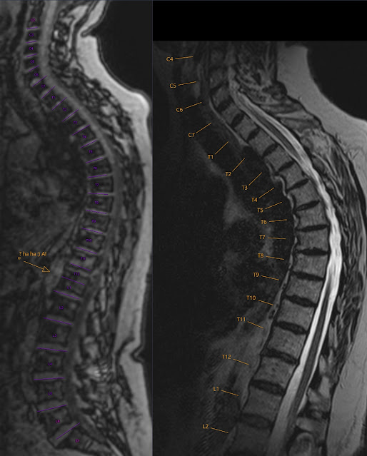
  As a funny post here example of counting spinal levels by AI (Artificial Intelligence - purple) vs Human (me - orange). :)

Music Staff on MRI Lumbar Spine - fun

Image
As a new hobby project I recently took on some digital piano and synthesizer training. Part of it is to learn to read the notes. And somehow I keep on seeing five lines of staff even om MRI images :) ! This localizer incidentally even shows the grand staff . :)  You can see a little annular tear in a place of previous disk herniation.  The numbering of lumbar levels is often a matter of discussion. Previous exam has called the lowest lumbar vertebra as L5. I used the same numbering but in fact the vertebra that is called L5 on this image is a transitional vertebra that is partially sacral vertebra - so it could be called S1. I made a note on my report for the clinician that in case of operation it is important to correlate the level of operation with fluoroscopy .  For correct numbering of spinal lumbar levels I look for proximal parts of ribs 12 on sagittal images and count from what I assume as L5 upwards. When I have the whole spine or CT, especially with coronal im...

Ventriculus Terminalis - Fifth Ventricle

Image
sag T2, ax T2, ax T1 C+, ax T1 C+ FS sag T2, T1 C-, T1 C+, T1 C+ FS As additional finding at MRI of Lumbar spine an oval cystic lesion with thin well demarcated wall without contrast enhancement located in conus medullaris. This is a  ventriculus terminalis also known as fifth ventricle . It represents developmental remnant and is believed to have no clinical significance. It has typical location and lack of enhancement differentiates it from other cystic lesions in the spine as for example hemangioblastoma.  See also this interesting article: G. Liccardo - Fifth ventricle: an unusual cystic lesion of the conus medullaris  

Spondylolysis

Image
When you see spondylolisthesis (minimal in our patient) you might suspect (usually  bilateral ) spondylolysis . This is often not the case as spondylolisthesis can also be due to displacement in the facet joints. However it is wise not to miss the spondylolysis. It is rather easy finding on CT - as you can see above. There is no problem judging spondylolysis from normal facet joints. However nowadays we mainly perform MRI for spinal problems. On MRI it is not that easy to find spondylolysis. This is mostly due to  more horizontal angle of the fractures compared to vertical angle of the normal facet joints as seen on transversal images. The sagittals are not always that helpful as well. The clue is that spondylolysis looks like 'additional joint' that should not be there. Note on the images above spondylolysis as well as normal facet joint with cartilage seen on the last image (long arrow). This is the same patient as shown on CT therefore it is good to compare those ...