Partial Double Internal Jugular Vein
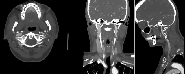
The above CT Angiography (CTA) shows Partial Double Internal Jugular Vein on the left side that starts at C2 level and continues to jugular foramen. Note normal dominant right internal jugular vein. This is a rare anatomic variant. I suppose we are going to see it more often as the use of CTA increases.
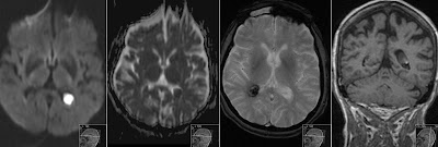
The same patient presents with a Hypoplastic Transverse Sinus on the left side and normal dominant on the right. This is a common anatomic variant.


