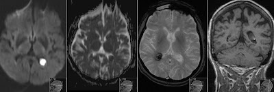
Late Subacute Hemorrhage on DWI
Axial susceptibility weighted image (SWI) shows hemorrhage in the right inferior frontal gyrus with hypointense rim consistent with hemosiderin and central hyperintensity due to extracellular methemoglobin. The high signal intensity is also seen on the coronal nonenhanced T1. Diffusion weighted image (DWI) shows high signal and low on the corresponding ADC map which is consistent with restricted diffusion due to extracellular methemoglobin. There is also focal hemosiderin deposit in the left parietal lobe - shown on SWI, as well as many other smaller hemosiderin foci (not shown). Patient is suspected for amyloid angiopathy or multiple cavernomas.


