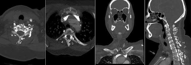
Contrast in Vertebral Venous Plexus
There is increased contrast in the vertebral venous plexus of the cervical spine. This is due to entrapment (positional exacerbated) of the left brachocephalic vein between aorta and sternum. This resulted in decreased (delayed) arterial contrast during this Carotid CTA.


