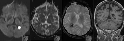
Cavum Velum Interpositum on MRI
Incidental finding of Cavum Velum Interpositum (CVI) that is seen as triangular shaped CSF space between lateral ventricles, over thalami, below fornices.
CVI is most posteriorly located - that is contrary to anterior and middle location of Cavum Septi Pellucidi (CSP) and Cavum Vergae (CV) as shown on this very good scheme from Wikipedia.
You might also check my previous post showing Cavum Velum Interpositum on CT.


