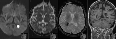
Sturge Weber CT
Non-contrast CT showing cortical calcifications and atrophy in the left occipital lobe in a patient with Sturge-Weber Syndrome. Due to leptomeningeal angiomatous venous plexus there are dystrophic cortical changes. In microscopy study those patients have a plexus of multiple small thin-walled telangiectatic capillaries or venules in the subarachnoid space between pia and arachnoid membranes. Diminished venous cortical draining causes cortical atrophy that can be seen on the second and third image. Cortical calcifications have "tram-track" pattern - see arrows fourth image. There is no significant contrast enhancement (not shown). Also enlarged ipsilateral sinuses are part of the intracranial findings - see FS on the first image.


