Basilaris Thrombosis

Note high density in the cranial part of the basilar artery (BA). First image showing normal density in the part without thrombus, the second transversal image more cranial showing thrombus, that is also visible on sagittal image. Normally basilar artery can be of higher density, however in this case you can see difference in various parts of the artery. There is also clinical information that plays significant role in diagnosis.

CT Angiography (CTA) confirms diagnosis of Basilar Artery Thrombosis (Basilaris Thrombosis) showing lack of contrast in the apical part of the basilar artery.

Patient was transferred immediately to Angiography Lab for selective catheterization and thrombolysis. Image on the left showing occlusion of BA and image on the right showing excellent recanalization after thrombolysis.
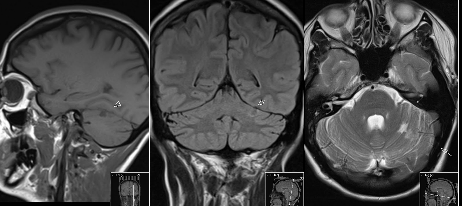
Follow up MRI has shown only a small cortical infarct in the upper parts of cerebellum as rest after the incident.

Also in favor for this patient was presence of both posterior communicating arteries, as shown above on the follow up ToF MR Angiography (MRA).


