Neurofibromatosis 1

This patient is known with Neurofibromatosis Type 1 (von Recklinghausen Disease). Note enlarged optic nerves on coronal T2, as well as thickened optic chiasm on coronal T1 with Gadolinium and thickened optic tracts on transversal T1 with Gadolinium. This is due to Optic Glioma. There is very little, almost no contrast enhancement. Last image, transversal T2, showing tumor infiltration in the optic tract as well as Focal Areas of Signal Intensity (FASI) - that show no enhancement.
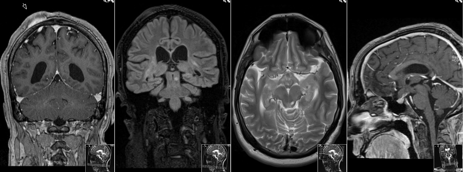
Also note enhancing Subcutaneous Neurofibroma, more FASI and how the thickened optic chiasm looks on sagittal T1.


