Herpes Simplex Encephalitis
6 months old child diagnosed with Herpes Simplex Encephalitis (HSE). CT showing low density areas in both temporal lobes. This finding is difficult on CT due to artefacts often present in middle cerebral fossa. Clue here is the coronal reconstruction showing low density in the lower parts of the basal ganglia.
MR in acute phase confirms diffusion restriction bilateral in the lower parts of the basal ganglia as well as oedema seen on T2.
T2 and FLAIR show diffuse oedema in the lower parts of basal ganglia on both sides.
Follow up MR about 3 months later showing on FLAIR and T1 extensive atrophy and gliosis in the temporal lobes.
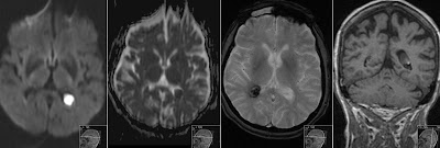
Axial SWI showing extensive hemorrhagic changes (hemosiderin) in the HSE damaged regions. Note extensive encephalomalacia involving temporal lobes and lower parts of basal ganglia.
Teaching point here is to have an extra careful look at CT images that are mostly performed as first line of examination, especially look for oedema and low density in both temporal lobes, lower parts of basal ganglia and the limbic system. It is recommended to perform MR study for further diagnosis. In older patients infarction is the main differential diagnosis. HSE is mostly bilateral and often asymmetric.


