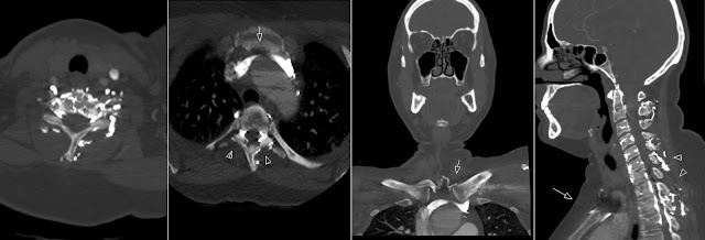
Posts

Showing posts from July, 2010

Skull Fracture Line

Image
This very thin skull fracture line without dislocation can be easily missed. It is hard to find on the axial images, well seen on the sagittal projection and easy to find on the 3D reconstructions. So my simple radiology tip is: "Look at all images".

PRES

Image
PRES - Posterior Reversible Encephalopathy Syndrome. Patient after two kidney transplantations that presents with seizures. Initial CT without contrast shows bilateral hypodensities cortical and in subcortical white matter in the parietooccipital regions. Initial MR (transversal and coronal T2 and axial FLAIR) shows signal abnormalities in the cortical and subcortical regions mostly occipital and parietal as well as frontal. Changes are believed to represent vasogenic edema. Follow up MR ten days later shows decrease of the pathological high signal changes (edema) with some residue signal abnormalities still visible.

Lissencephaly

Image
One year old child with microcephaly, psychomotor retardation and deletion on chromosome 17. Transversal T2, IR, coronal FLAIR and parasagittal T1 show decreased number of sulci as well as clearly thickened gyri. Case of Lissencephaly - "smooth brain" with Pachygyria - "broad gyri". Images from 3T MRI scanner.

Falx Meningioma

Image
Large Falx Meningioma is an easy find on contrast enhanced CT. However note how isodense is the tumor on the non contrast enhanced CT. Also due to it's cranial location it can be missed on axial images. Therefore look carefully at those upper slices on the axial series and it is highly recommended to include coronal and sagittal reformations as standard evaluation protocol.

Cerebral Venous Sinus Thrombosis

Image
CT Venography (CTV) Maximum Intensity Projection (MIP) images show contrast filling defects in the right sigmoid sinus, right transversal sinus and superior sagittal sinus. Extensive Cerebral Venous Sinus Thrombosis. Note prominent collateral circulation in the posterior fossa on the right side.

Contrast in Vertebral Venous Plexus

Image
There is increased contrast in the vertebral venous plexus of the cervical spine. This is due to entrapment (positional exacerbated) of the left brachocephalic vein between aorta and sternum. This resulted in decreased (delayed) arterial contrast during this Carotid CTA.

Moyamoya

Image
Coronal contrast enhanced T1 shows increased number and size of the lenticulostriate arteries "puff of smoke" that represent collateral circulation as a consequence to the tapering of the great intracerebral vessels. This is a case of Moyamoya. Note reduced caliber of the left MCA shown on the ToF MRA images. Also note on the lateral MIP from the ToF MRA a defect in the basilar artery flow with multiple collateral vessels.

Mandibula Fracture

Image
Undislocated mandibula fracture. Note that fracture goes through mental foramen and apex of the root of tooth 34.

Developmental Venous Anomaly

Image
Contrast enhanced T1 images show large Developmental Venous Anomaly (DVA) in the cerebellum on the right side as incidental finding. Large vein is draining into the sinus. Another incidental finding is a small meningioma parietal on the right.

Tolosa-Hunt Syndrome

Image
Tolosa-Hunt syndrome is clinically a painful ophthalmoplegia cused by inflammatory lesion of the cavernous sinus that is steroid responsive. Pathologically this process is similar to orbital pseudotumor. Above axial non contrast enhanced and axial, coronal and sagittal contrast enhanced CT shows a lesion extending from the cavernous sinus into the orbit through the superior orbital fissure. According to the literature CT is often normal. This case however proves value of the CT. MR is the investigation of choice in order to exclude other pathology in this region. See below reconstruced images. See also: AJR Teaching File: Cavernous Sinus Mass in a Woman Presenting With Painful Ophthalmoplegia - Asim K. Bag