Diffusion Weighted Imaging - MRI
Diffusion Weighted Imaging (DWI) is a fast and very usefull MRI sequence that should be included in every brain scan. Quality is good on scanners with 1.5 T and more.
When you set the images in such order: DWI, ADC and T2 (b0) the configuration of signal: white - black - white indicates Restricted Diffusion.


See also case of Late Subacute Hemorrhage on DWI with restricted diffusion due to extracellular methemoglobin.

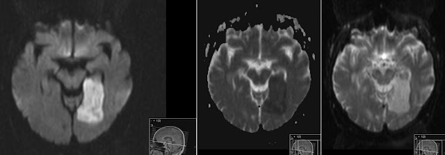
PCA Infarct
Above: subacute arteria cerebri posterior infarct in the left occipital lobe. Note restricted diffusion (high signal) on the isotropic DW image (the one with black CSF). Note black corresponding area on the ADC map (image with white CSF and "spots" around the brain). High signal on the last b0 image that is confirming T2 signal of the infarct.When you set the images in such order: DWI, ADC and T2 (b0) the configuration of signal: white - black - white indicates Restricted Diffusion.

Small (lacunar) thalamus infarct
Above: also infarct. This time a lacunar infarct in the left thalamus showing Restricted Diffusion on the DWI images.
Glioblastoma
Above: Glioblastoma tumor in the left temporal lobe showing rim enhancement on post contrast T1 and central necrosis. The necrotic part of the tumor shows No Restriction - same signal as CSF. Black - white - white.

Abscess
Above: Abscess in the right frontal lobe showing Restricted Diffusion.
Diffusion is esspecially helpful in distinguishing abscess from necrotic parts of the tumor.

Metastasis
Above: Lung cancer metastasis showing No Restricted diffusion - just like necrotic parts of the tumor.
Multiple metastasis
Above: Multiple metastasis of the Small Cell Lung Cancer. Those are mostly solid lesions and show Restricted Diffusion. They do not have so much necrotic fluid inside.

Meningioma
Above: large extra axial solid tumor Meningioma showing Restricted Diffusion.


