Intracranial Hemorrhage on MRI
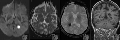
Case of a 2 days old neonate with bilateral subdural hematomas, mainly infratentorial. Note high signal on the coronal T1, low on GRE, high signal with level on T2 and same level on T1 sequences. Signal characteristics represent early subacute hematomas with methemoglobin still in the red blood cells. I would like to use this case as a reminder of signal changes of intracranial hematomas on MRI.
The table above shows how we stage (name) hematomas according to time. Important observation is that the very early (hyperacute) hematomas contain Oxyhemoglobin and are difficult to see (isodense to brain) on T1 sequences. Same with Deoxyhemoglobin. Then after about 3 days we start to see high signal of Methemoglobin on T1. That continues to be high on T1 even when Methemoglobin is released from the hemolyzed Red Blood Cells, but then we start to see it as high even on T2. Late remains of the hemorrhage on MR can be seen as a rim of Hemosiderin deposits - that is just black. Gradient Echo (T2*) (GRE) sequences show hemorrhage as black since it is a sort of susceptibility artefact. It also exaggerates the volume of bleeding ("blooming artefact").
You may also check:
Neonatal Intraventricular Hemorrhage
Hemorrhagic Choroid Plexus Cyst
Late Subacute Hemorrhage on DWI
Hemorrhagic Brain Metastases


