Thalamic Infarct
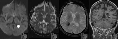
First image shows Acute Lacunar Thalamic Infarct on the right side with corresponding second image showing prior study 11 hours before, that also depicts acute infarction.
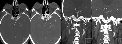
CTA shows occlusion of the P1 segment of the right Posterior Cerebral Artery (arrows). You can also (with some difficulty) depict a thromb in this segment (arrowheads).
Lacunar Thalamic Infarcts are quite common and often accompanied with Posterior Cerebral Artery territory infarcts. In fact PCA infarcts are detected more often. However isolated thalamic infarcts are also seen. The clue here is vascular supply to the thalami. It comes mainly from P1 and P2 segments of the Posterior Cerebral Arteries. Therefore radiologists and neurologists should pay attention to posterior circulation in case of suspected or detected thalamic infarcts. Patients with such infarcts present with specific neurologic findings.
Excellent review of vascular supply can be found in the following articles:
Jeremy D. Schmahmann - Vascular Syndromes of the Thalamus
Young-Mok Song - Topographic patterns of thalamic infarcts in association with stroke syndromes and aetiologies
You can also check my previous post showing examples of thalamic and PCA infarcts on MRI
Diffusion Weighted Imaging - MRI


