Posts

Showing posts from March, 2011

CSF Leakage - Intracranial Hypotension

Image
Young woman two weeks after partus followed by spinal pain and stiffness in the neck. Sagittal T2 and contrast enhanced T1 show: sagging of the midbrain as well as cerebellar tonsils, enlarged dural sinuses and hypophysis, steep tentorium. Axial contrast enhanced T1 showing increased dural enhancement and slightly increased subarachnoidal space seen on coronal T2. (BTW: Also note nicely depicted corticospinal tracts on the coronal T2.) Coronal contrast enhanced T1 shows increased dural enhancement as well as sagging of the cerebellar tonsils. Findings strongly suggest Intracranial Hypotension as a consequence of the reduced Cerebro Spinal Fluid (CSF) pressure due to possible CSF leakage associated with lumbar puncture (LP). Differential diagnosis are: 'normal' dural enhancement after LP, as well as meningitis. However meningitis tends to show more subarachnoidal than dural enhancement. See also other case of Intracranial Hypotension from my blog.

Optic Nerve Sheath Meningioma

Image
Contrast enhanced CT showing well defined, homogeneous intra orbital tumor. There is relation of the tumor with optic sheath and rather deep location in the orbital apex. This is most likely an  Optic Nerve Sheath Meningioma .

Just Looking!

Image
Amusing T2 slice from 3T MRI scanner showing right sided fixed position of the lenses. Perhaps our patient saw something interesting in the scanner? :) For more fun radiology images check older posts in Fun label.

Hemorrhagic Choroid Plexus Cyst

Image
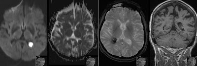
Transversal Diffusion Weighted sequence shows high signal in the left choroid plexus with corresponding low signal on ADC (restricted diffusion). Finding represents acute Hemorrhagic Choroid Plexus Cyst . It is also known as Choroid Plexus Xanthogranuloma . Gradient Echo sequence (third image) shows low signal in the right choroid plexus representing hemosiderine deposits after previous hemorrhage. Coronal T1 shows intermediate to high signal in the plexus cyst. Note also low signal in the right plexus on T2 (old hemorrhage) and high signal on FLAIR in the left plexus (acute hemorrhage). Choroid Plexus Cysts are very common normal finding. Those often show restricted diffusion. However in our case of patient with hypertension this finding is of interest to the clinicians as a requirement to more closely monitor patient's blood pressure. There were no signs of intraventricular hemorrhage.

AV Hemodialysis Shunt Thrombosis

Image
CTA (Computed Tomography Angiography) showing nice 3D rendering of the AV Hemodialysis Shunt Thrombosis (purple) in the cephalic vein with dilatation of the distal part of the cephalic vein. The Arterio-Venous shunt is open. Source images of the same investigation show that there is partially blood flow around thrombus and dilated distal part of the cephalic vein. Important note: CTA is not the investigation of choice for the suspected hemodialysis shunt thrombosis - Color Doppler Ultrasound is the investigation that should be performed first. This case was investigated in order to provide surgeon with anatomic topography of the anastomosis and in this way replacing angiography.

Struma

Image
3D rendering of the CT Angiography showing nicely a large struma displacing trachea without deforming trachea lumen. Eye Candy.

Epidermoid Cyst

Image
Note high signal on DWI of this cystic process in the left cerebellopontine angle. This is Epidermoid Cyst showing lobulated contour and mass effect on brainstem. On ADC signal resembles brain parenchyma. It shows slightly decreased signal on T2 and no contrast enhancement. Sagittal T1 and coronal FLAIR show signal of Epidermoid to be slightly higher than CSF. Somehow it resembles signal characteristics of Cholesteatoma that is also keratin based cystic inclusion process.