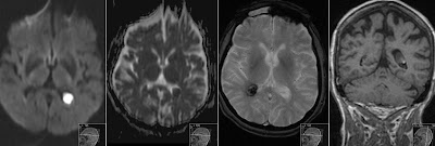
Epidermoid Cyst
Note high signal on DWI of this cystic process in the left cerebellopontine angle. This is Epidermoid Cyst showing lobulated contour and mass effect on brainstem. On ADC signal resembles brain parenchyma. It shows slightly decreased signal on T2 and no contrast enhancement.
Sagittal T1 and coronal FLAIR show signal of Epidermoid to be slightly higher than CSF.
Somehow it resembles signal characteristics of Cholesteatoma that is also keratin based cystic inclusion process.
Sagittal T1 and coronal FLAIR show signal of Epidermoid to be slightly higher than CSF.
Somehow it resembles signal characteristics of Cholesteatoma that is also keratin based cystic inclusion process.


