Cavernoma - Cavernous Angioma
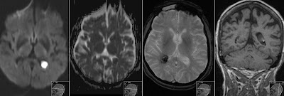
CT without contrast shows hyperdense lesion in the left occipital lobe. T2 shows hemosiderin deposits without surrounding edema. T1 with contrast shows no enhancement. The most characteristic however is the last image: T2* Gradient Echo showing susceptibility blooming artifact - that is larger than lesion itself.


