Chronic Subdural Hematoma
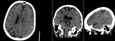
Above unenhanced CT shows a classic case of bilateral Chronic Subdural Hematomas (cSDH). Note that hematomas are relatively isodense with brain tissue. Due to this isodenisty cSDH can sometimes be missed. Also note fresh blood in the cSDH on the patient's left side. Coronal image nicely depicts subarachnoidal space. The sagittal image is off center to the left side of the patient.


