Hemorrhagic Brain Metastases
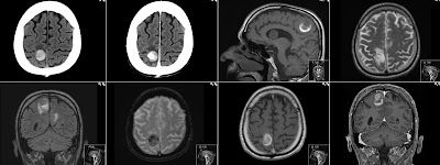
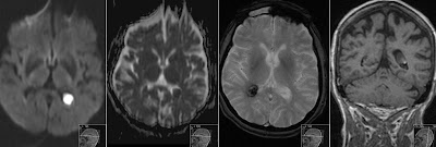
This patient has multiple hemorrhagic brain metastases from the lung cancer. Note hyperdense metastasis in the right parietal lobe on the first unenhanced CT image. CT with contrast shows vivid enhancement of this tumor. Also note hypodense surrounding edema. Sagittal unenhanced T1 shows rim of high signal around metastasis that is caused by methemoglobin from the subacute hemorrhage. Also note intraventricular metastasis. Axial T2 shows surrounding edema. Coronal FLAIR shows edema, also from other metastasis location. Axial T2* Gradient Echo shows black signal due to hemosiderine. Axial T1 with Gd shows enhancement as well as coronal T1 Gd. In case of this patient the active hemorrhage from some of the metastasis have caused symptoms.


