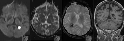
Oculomotor Infarct
This 40 years old patient has experienced acute oculomotor nerve paresis. A 1.5T MRI has revealed a micro infarct (about 2mm) in location of the oculomotor nerve nucleus in the mesencephalon. Note high signal on DWI (B1000), low signal on ADC and high on T2 (B0) of the Diffusion Weighted Images (DWI) and coronal FLAIR. This case illustrates how sensitive can DWI be even in small infarcts.
Above drawing courtesy of Bartleby.
Fig. 710 Anatomy of the Human Body by Henry Gray
This is also a great opportunity to remind the location of the oculomotor nerve nucleus in mesencephalon (8'). Note that mesencephalon here is "facing down" compared to MRI. Well seen structures on the MRI are Red Nucleus, Substantia Nigra and Cerebral Aqueduct.

Cranial Nerves Nuclei
Drawing titled: Brain stem human sagittal section - by Patrick J. Lynch, medical illustrator
Courtesy Wikipedia
Also note location of the other Cranial Nerves Nuclei in the brainstem that are nicely depicted in the above drawing.
Above drawing courtesy of Bartleby.
Fig. 710 Anatomy of the Human Body by Henry Gray
This is also a great opportunity to remind the location of the oculomotor nerve nucleus in mesencephalon (8'). Note that mesencephalon here is "facing down" compared to MRI. Well seen structures on the MRI are Red Nucleus, Substantia Nigra and Cerebral Aqueduct.

Cranial Nerves Nuclei
Drawing titled: Brain stem human sagittal section - by Patrick J. Lynch, medical illustrator
Courtesy Wikipedia
Also note location of the other Cranial Nerves Nuclei in the brainstem that are nicely depicted in the above drawing.


