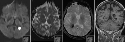
Congenital Cytomegalovirus Infection
Four weeks old neonate with biopsy proven Cytomegalovirus (CMV) hepatitis was investigated with MRI of the brain due to altered neurologic status. T2* GRE Gradient Echo image shows multiple small black dots bilateral periventricular that represent multiple small calcifications. FLAIR shows extensive diffuse signal abnormalities in the white matter. Transversal T2 TSE shows calcifications round occipital horn of the left lateral ventricle. Coronal T1 shows high signal intensity small dots infratentorial that also represent small calcifications. (Yes - calcifications can show as high signal on T1). Periventricular distribution of calcifications is characteristic for Congenital Cytomegalovirus (CMV) Infection. Those would be even better visible on CT and even on Ultrasound.
However MRI shows further characteristic findings. Note pathological structure of the gyri on the parasagittal and transversal T2 images corresponding with dysplastic cortex - polymicrogyria. Also note on the sagittal T2 and T1 images a hypoplastic thin corpus callosum as well as large cisterna magna.


