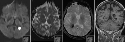
Epidermoid Cyst DWI
CT (not shown) has revealed a cystic structure in the left cerebellopontine angle with mass effect on the pons and cerebrellar peduncle. MR has shown that the structure has inhomogeneous slightly increased signal on FLAIR, cystic high signal on T2 and what is most characteristic has high signal on diffusion weighted (DWI) sequence. On ADC signal was very close to that of brain parenchyma. Also note the expansion of the mass into the Meckel cave seen on FLAIR (first image). This finding represents a classic Epidermoid Cyst.
You might also check another case of Epidermoid Cyst in this most common location.


