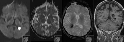
Sinus Thrombosis - 9 days old neonate - MRI
9 days old neonate. Mother known with sinus thrombosis during pregnancy. This is T1 without contrast showing high signal of methemoglobine in sinus rectus, superior sagittal sinus and cavernous sinus - representing massive blood clots of sinus thrombosis.
SWI sequence is showing low signal of hemosiderine deposits in central veins. T2 sequence is showing clot filled occipital part of sagittal sinus. ToF MRV (flow based venous angio) shows corresponding flow defects, same on MRV MiP reconstruction.
DWI and ADC-map showing ischemia in the basal ganglia, corpus callosum and corona radiata frontally. Those ischemic changes can be reversible in case of sinus thrombosis (venous infarcts). Major risk are the possible brain hemorrhages - that are fortunately not present in this case.
Teaching point is to look very carefully at non-contrast T1 of neonate for possible high signal changes representing methemoglobine products, not only in dural sinus but also in the subarachnoidal spaces for possible partus related subdural hematomas (SDH).
You can check my post about Intracranial Hemorrhage on MRI


