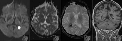
2 small abscesses
Immunocompromised patient presents with ring-enhancing subcortical expansivities with perifocal oedema. MRI was performed (not shown) suggesting 2 small abscesses. Patient was set on antibiotics.
Control MRI after one month shows on axial T1 without- and with contrast slightly decreased size of ring enhancing expansivities.
Both small abscesses are still enhancing.
There is still perifocal vasogenic edema seen on axial FLAIR and T2 weighted sequences. Note on T2 a well defined low signal rim of the abscesses.
Diffusion weighted imaging showing central diffusion restriction (high signal on DWI and low on ADC-map).
Numerous pathologic entities show as ring-enhancing lesions therefore it is important to include patient's history in evaluation. Here the biggest clue is restricted diffusion. However hemorrhage can also show restricted diffusion due to high protein content as well as rim of hemosiderin on T2.
You can also check my other cases related to this topic:
Abscess and Subdural Empyema
Diffusion Weighted Imaging - MRI
Late Subacute Hemorrhage on DWI


