CT Pelvimetry - Bäckenmätning
Below is a short summary of the protocol for Pelvimetry (Bäckenmätning in Swedish).

First a thick MIP is reconstructed on the scanner that covers whole pelvis. A line is drawn on the scanner that is later used for calibration on PACS workstation.

Calibration of the previously measured line.

On the above MIP the Transversal Inlet Diameter (Transverstingångsmått) is measured as the largest transversal distance in the pelvic inlet. As well as Intertubar Diameter (Intertubaravstånd) as distance between ischial tubers.

On sagittal reconstruction measure Sagittal Inlet Diameter (Sagittellt ingångsmått) as shortest distance between posterior part of symphysis pubis and promontorium (S1). As well as Sagittal Outlet Diameter (Sagittellt utgångsmått) as shortest distance between posterior part of symphysis pubis and fist mobile joint between sacrum and coccyx.

On axial reconstruction (see topogram for level) measure Interspinal Diameter (Interspinal avstånd) as shortest distance between ischial spines.

In your final report group the measurements in two groups:
Sagittal Inlet Diameter
Transversal Inlet Diameter
Sagittal Outlet Diameter
Interspinal Diameter
Intertubar Diameter
Some obstetrics departments like to see the summation of the values for the inlet and outlet measurements.
Below some more examples:

Sagittal Inlet Diameter - note the shortest distance to measure.

Pelvic MIP showing Transversal Inlet Diameter measurement.
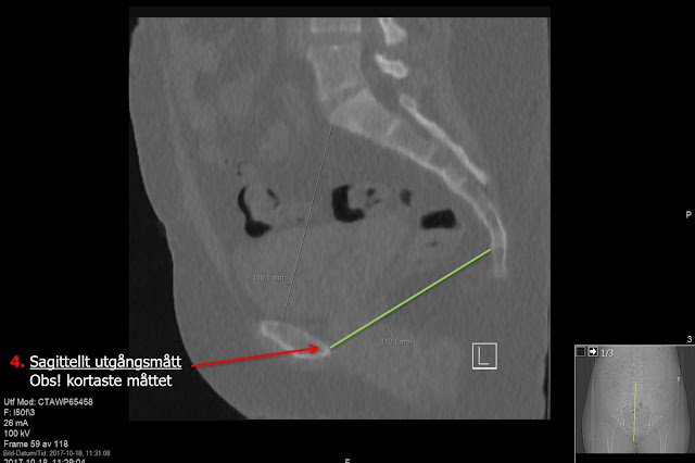
Sagittal reconstruction for Sagittal Outlet Diameter - measure the shortest between posterior part of symphysis pubis and fist mobile joint between sacrum and coccyx.

Axial reconstruction for Interspinal Diameter - again measure the shortest distance between ischial spines.

On MIP measure Intertubar Diameter - measure from the central parts of the ischial tubers.
Your final report could look like this (if you report in Swedish :)

With Great Thanks for lending me the images to Dr. Rahideh Azadeh - radiology resident in Sweden!

First a thick MIP is reconstructed on the scanner that covers whole pelvis. A line is drawn on the scanner that is later used for calibration on PACS workstation.

Calibration of the previously measured line.
On the above MIP the Transversal Inlet Diameter (Transverstingångsmått) is measured as the largest transversal distance in the pelvic inlet. As well as Intertubar Diameter (Intertubaravstånd) as distance between ischial tubers.

On sagittal reconstruction measure Sagittal Inlet Diameter (Sagittellt ingångsmått) as shortest distance between posterior part of symphysis pubis and promontorium (S1). As well as Sagittal Outlet Diameter (Sagittellt utgångsmått) as shortest distance between posterior part of symphysis pubis and fist mobile joint between sacrum and coccyx.

On axial reconstruction (see topogram for level) measure Interspinal Diameter (Interspinal avstånd) as shortest distance between ischial spines.

In your final report group the measurements in two groups:
Sagittal Inlet Diameter
Transversal Inlet Diameter
Sagittal Outlet Diameter
Interspinal Diameter
Intertubar Diameter
Some obstetrics departments like to see the summation of the values for the inlet and outlet measurements.
Below some more examples:

Sagittal Inlet Diameter - note the shortest distance to measure.

Pelvic MIP showing Transversal Inlet Diameter measurement.
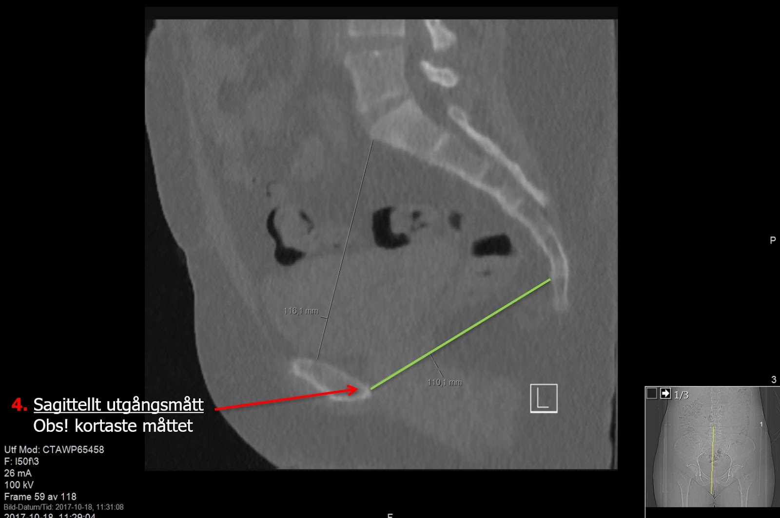
Sagittal reconstruction for Sagittal Outlet Diameter - measure the shortest between posterior part of symphysis pubis and fist mobile joint between sacrum and coccyx.

Axial reconstruction for Interspinal Diameter - again measure the shortest distance between ischial spines.

On MIP measure Intertubar Diameter - measure from the central parts of the ischial tubers.
Your final report could look like this (if you report in Swedish :)

With Great Thanks for lending me the images to Dr. Rahideh Azadeh - radiology resident in Sweden!


