Posts

Showing posts from October, 2009

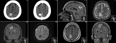
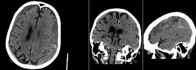
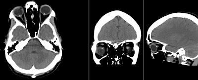
Anterior Communicating Artery Aneurysm Rupture

Image
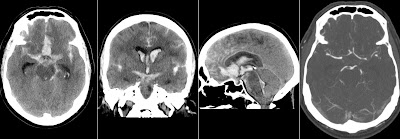
Massive hemorrhage from ruptured Anterior Communicating Artery (Acom) aneurysm with blood in basal cysterns, subarachnoid space and ventricular system. Note on the sagittal image delineation of the third ventricle, aqueduct and fourth ventricle with blood. CT Angiography (CTA) shows 3mm Acom aneurysm. Note dilatation of the ventricular system, particularly temporal horns on the first image. Intraventricular hemorrhage is causing acute hydrocephalus.

Dental Caries on CT

Image
This patient has extensive Dental Caries in the right 6th lower tooth (46). Note also status post extraction of the 6th lower tooth on the left (36) and artifacts from fillings in upper teeth (coronal). This is an incidental finding on CT after head trauma that shows nicely the capability of this technique.

Hemorrhagic Brain Metastases

Image
This patient has multiple hemorrhagic brain metastases from the lung cancer. Note hyperdense metastasis in the right parietal lobe on the first unenhanced CT image. CT with contrast shows vivid enhancement of this tumor. Also note hypodense surrounding edema. Sagittal unenhanced T1 shows rim of high signal around metastasis that is caused by methemoglobin from the subacute hemorrhage. Also note intraventricular metastasis. Axial T2 shows surrounding edema. Coronal FLAIR shows edema, also from other metastasis location. Axial T2* Gradient Echo shows black signal due to hemosiderine. Axial T1 with Gd shows enhancement as well as coronal T1 Gd. In case of this patient the active hemorrhage from some of the metastasis have caused symptoms.

Toyota iQ

Image
Today I took Toyota iQ as replacement for my car that I left for service. It was 1.0 L 3 cylinder benzine with manual transmission. I normally drive a 4x4 2005 Land Cruiser 3.0 L 4 cylinder diesel with automat. So it was quite interesting change for me. The reason was that I like the iQ concept: make it small on the outside and large on the inside . iQ is practically square. In white it looks like a washing-machine. But most of the time we spend sitting inside the car and not looking at it’s outside. In Europe the road tax for heavy, diesel cars is very high. A little and economic car seems really interesting nowadays. iQ is a perfect car for one person living in the city. It’s easy to park and not obtrusive. I like the looks of iQ. Inside it feels quite roomy (for one). Seats are a bit too hard, however allowing a comfortable adjustment. (After my second test a few weeks later I must admit that seats are not that comfortable. That is sad as the price of this car is rather high in

Wallenberg Syndrome - Lateral Medullary Syndrome

Image
DWI (b1000) image shows restricted diffusion indicating right sided dorsal lateral medulla oblongata infarct. Corresponding ADC map image shows low signal and axial T2 and coronal FLAIR show high signal in this Posterior Inferior Cerebellar Artery (PICA) territory infarct. This case shows how sensitive is MRI in detecting even little infarcts. Also the eponym is worth noting in case of exams ;).

Central Pontine Myelinolysis

Image
Axial T2 shows increased signal centrally in the pons. Axial, sagittal and coronal T1 with Gd show enhancement of those regions with sparing of the corticospinal tracts. The term Central Pontine Myelinolysis (CPM) is now preferably named as Osmotic Demyelination Syndrome (ODMS) due to common association with rapidly corrected hyponatremia. As a radiologist I prefer the CPM name as it is more anatomic specific about the location of the findings. However do not forget the term ODMS when looking for it in the textbook index.

iPhone on CT Scanogram

Image
I always recommend checking the Scanogram on every CT. It can reveal prior operations, foreign bodies - that can be of importance for MRI screening. One can also appreciate patient's general constitution. Sometimes as in this case it can reveal what type of mobile device the patient is using and that he needs glasses to read from it ;).

Chronic Subdural Hematoma

Image
Above unenhanced CT shows a classic case of bilateral Chronic Subdural Hematomas (cSDH). Note that hematomas are relatively isodense with brain tissue. Due to this isodenisty cSDH can sometimes be missed. Also note fresh blood in the cSDH on the patient's left side. Coronal image nicely depicts subarachnoidal space. The sagittal image is off center to the left side of the patient.

H1N1 Swine Flu causing ARDS

Image
Above Chest X-ray shows patchy densities covering mostly basal and medial parts of both lungs. This is a case of Acute Respiratory Distress Syndrome (ARDS) as a complication of the H1N1 virus infection. This virus on contrary to common flu virus has capacity to bind to the cellular receptors that are situated deep in the lungs. Therefore can cause massive pneumonia very rapidly. On contrary common flu virus binds mostly to the receptors in the upper respiratory tract. Pandemic flu can infect cells deep in the lungs, says new research

Candy Under the Tongue on CT

Image
Above CTA shows hyperdense (450 HU) oval structure under the tongue. Imaging is with iv contrast so one could wonder if this is a hyperdense or enhancing lesion? However it is really just a candy that the patient has had under her tongue during examination. Also note the artifacts from dental fillings. Sometimes is worth to angulate the head to avoid those artifact in the regions of interest. There are even some articles about foreign bodies in oral cavity: McDermott M - What's in your mouth? The CT appearance of comestible intraoral foreign bodies. Towbin AJ - The CT appearance of intraoral chewing gum.

Dilated Perivascular Spaces in the Brain

Image
Above images (tra T2, tra FLAIR, cor T2 and sag T1) show enlarged Perivascular Spaces (PVS) in the right frontal lobe of the elderly patient with dementia. Note that signal follows that of the CSF (see FLAIR). Moreover note moderate general cortical atrophy. PVS, also known as Virchow-Robin spaces are considered normal finding although perhaps in the future we should know more about the exact patophysiology and possible relations till dementia and other conditions.

My Dogs: Alf and Nikki

Image
Alf is about 2.5 years old German Shepherd male - born in Holland. Nikki is about 2.5 years old Siberian Husky female - born in Sweden. They both love each other. Husky is a no-nonsense dog. No barking, only when playing. Natural hunter. Left without lead will hunt even large animals. Fantastic for mountain-biking and outdoor sports. Can not be alone. Needs companion. Husky is attached but does not show it much. On contrary German Shepherd is very attached and will follow you even in the toilet. Husky does not make a watch dog. Is friendly to all. On the other hand German Shepherd will guard the house and surroundings. Alf can be aggressive if frightened, but is very sensitive and will not kill at once. Will ask for permission first! ;-) Kidding!  

Partial Double Internal Jugular Vein

Image
The above CT Angiography (CTA) shows Partial Double Internal Jugular Vein on the left side that starts at C2 level and continues to jugular foramen. Note normal dominant right internal jugular vein. This is a rare anatomic variant. I suppose we are going to see it more often as the use of CTA increases. The same patient presents with a Hypoplastic Transverse Sinus on the left side and normal dominant on the right. This is a common anatomic variant.

Neonatal MRI Faces

Image
This is not a Thai Buddist temple guard. Above image is a coronal T2 of the neonate. Note normal teeth buds in the maxilla. Also low signal from lenses. This is not a cute monkey. Not even Homer Simpson. Above image is a coronal T1 of the neonate. Note normal fat in chicks.

Diffusion Weighted Imaging - MRI

Image
Diffusion Weighted Imaging (DWI) is a fast and very usefull MRI sequence that should be included in every brain scan. Quality is good on scanners with 1.5 T and more. PCA Infarct Above: subacute arteria cerebri posterior infarct in the left occipital lobe. Note restricted diffusion (high signal) on the isotropic DW image (the one with black CSF). Note black corresponding area on the ADC map (image with white CSF and "spots" around the brain). High signal on the last b0 image that is confirming T2 signal of the infarct. When you set the images in such order: DWI, ADC and T2 (b0) the configuration of signal: white - black - white indicates Restricted Diffusion. Small (lacunar) thalamus infarct Above: also infarct. This time a lacunar infarct in the left thalamus showing Restricted Diffusion on the DWI images. Glioblastoma Above: Glioblastoma tumor in the left temporal lobe showing rim enhancement on post contrast T1 and central necrosis. The necrotic

Optic Disc Drusen

Image
Bilateral calcifications in optic discs shown on this non contrast CT. Sometimes it can be incidental finding. This young patient had visual symptoms. I suppose we can see ODD more often due to thin slices of the CT we use. Interesting review article about Optic Disc Drusen: Optic Disk Drusen by Auw-Haedrich C, Staubach F, Witschel H. Surv Ophthalmol. 2002 Nov-Dec;47(6):515-32. PMID: 12504737 Also Wikipedia entry: Optic Disc Drusen

Cavernoma - Cavernous Angioma

Image
CT without contrast shows hyperdense lesion in the left occipital lobe. T2 shows hemosiderin deposits without surrounding edema. T1 with contrast shows no enhancement. The most characteristic however is the last image: T2* Gradient Echo showing susceptibility blooming artifact - that is larger than lesion itself.

Color art in CT Perfusion images of the brain.

Image
Here some nice colors of the CT Perfusion images of the brain. Showing: Mean Transit Time (MTT) - how fast the blood flows through the brain, Cerebral Blood Flow - how much blood flows in a specific period (ml of blood/100g tissue/min), and Cerebral Blood Volume - how much blood is in a volume of tissue. Excellent pdf article on CT Perfusion of the Brain from Martin Wiesmann is here:  CT Perfusion of the Brain - Martin Wiesmann  
Image
Hi, Welcome to my blog.